GIN Home
Ichneumonid Morphology
Subfamily Key
Lists of World Genera
Acaenitinae
Brachycyrtinae
Collyriinae
Lycorininae
Ophioninae
Poemeniinae
Rhyssinae
Stilbopinae
Xoridinae
Ichneumonid Morphology
The morphological terminology is mostly that of Townes (1969). The following treatments of Hymenoptera morphology
are recommended to the interested student: Michener (1944), Bohart & Menke (1976), and Huber & Sharkey (in: Goulet & Huber, 1993).
Mesosoma and metasoma are used to refer to the apparent thorax and abdomen, respectively. MS1 is used for the first metasomal segment (second true abdominal segment). T1, T2, etc., are used for the first metasomal and subsequent tergites; S1, S2, etc., are used for the first metasomal and subsequent sternites.
In describing the anterior, posterior, dorsal, and ventral aspects of leg segments, the legs are imagined as stretched out horizontally, at right angles to the body. This is an unnatural position but is the one used in taxonomic descriptions.
The nomenclature of wing veins and cells is based upon Ross (1936) and Mason (1990). Veins are divided into abcissae, or sections, by their intersection with other veins. For example, the second abcissa of fore wing vein 1A (between 1cu-a and 2cu-a) is written as "2/1A."
In the figure captions below, terms used by Townes are in parentheses. In addition, there is a list of expanded definitions of special terms frequently used in the keys and descriptions.
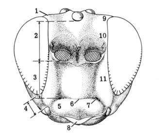
HEAD OF AN ICHNEUMONID, ANTERIOR AND POSTERIOR VIEWS
|
|
|
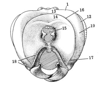
1, 13. Vertex 2. Supra-antennal area 3. Supraclypeal area 4. Malar space (= cheek) 5. Clypeus 6. Groove between face and clypeus 7. Anterior tentorial pit (= clypeal fovea) 8. Labrum |
9, 10, 11. Paraocular area 12. Genal orbit 14. Occiput 15. Foramen magnum 16, 17. Occipital carina (17 = genal carina) 18. Hypostomal carina (= oral carina) 19. Gena (= temple) |
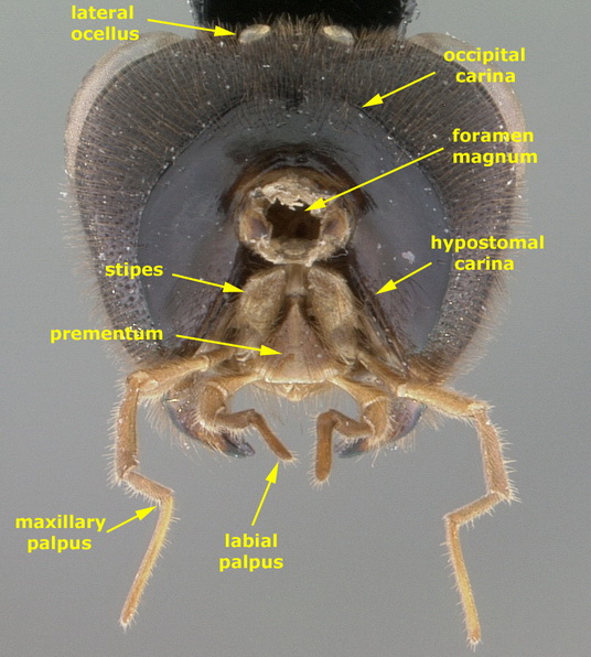
HEAD OF AN ICHNEUMONID (Saranaca apicalis), POSTERIOR VIEW SHOWING MOUTHPARTS
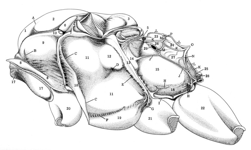
MESOSOMA OF AN ICHNEUMONID, LATERAL VIEW
|
1. Median lobe of mesoscutum 2. Lateral lobe of mesoscutum 1, 2. Mesoscutum 3. Scutellum 4. Metanotum (= postscutellum) 5. Hind margin of metanotum 6. Tegula 7. Subalar ridge (= subtegular ridge) 8. Collar |
8, 9, 10. Pronotum 10. Hind corner of pronotum 11, 12, 13, 18, 19. Mesopleuron 11, 12, 18, 19. Mesepisternum 12. Hypoepimeron (= speculum) 13. Mesepimeron 14. Dorsal division of metapleuron 15. Ventral division of metapleuron 16. Juxtacoxal area 17. Propleuron 18. Epicnecium (= prepectus) |
19. Mesothoracic venter 20. Fore coxa 21. Middle coxa 22. Hind coxa 23-28. Propodeum 23. First lateral area 24. Second lateral area 25. Third lateral area 26. First pleural area 27. Second pleural area 28. Third pleural area 29. Propodeal spiracle. |
|
A. Notaulus B. Epomia C. Epicnemial carina (= prepectal carina) D. Scrobe (= mesopleural fovea) E. Mesopleural groove F. Sternaulus G. Posterior transverse carina of mesothoracic venter H. Juxtacoxal carina I. Submetapleural carina J. Pleural carina |
K. Lateral longitudinal carina of propodeum L. Median longitudinal carina of propodeum M. Anterior transverse carina of propodeum (= basal transverse carina) N. Posterior transverse carina of propodeum (= anterior transverse carina) O. Propodeal apophysis or crest P. Costula [part of anterior transverse carina] |
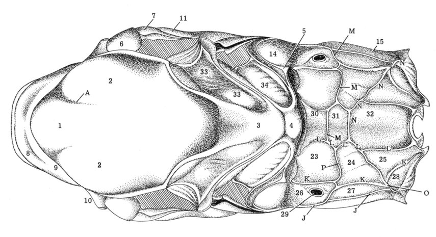
MESOSOMA OF AN ICHNEUMONID, DORSAL VIEW
|
1. Median lobe of mesoscutum 2. Lateral lobe of mesoscutum 1, 2. Mesoscutum 3. Scutellum 4. Postscutellum 5. Hind margin of metanotum 6. Tegula 7. Subalar ridge (= subtegular ridge) |
8. Collar 8, 9, 10. Pronotum 10. Dorsolateral corner of pronotum 11. Mesopleuron [mesepisternum] 14. Dorsal division of metapleuron 15. Ventral division of metapleuron 23-32. Propodeum 23. First lateral area 24. Second lateral area |
25. Third lateral area 26. First pleural area 27. Second pleural area 28. Third pleural area 29. Propodeal spiracle 30. Basal area 31. Areola 32. Petiolar area 33. Axillary trough of mesonotum 34. Axillary trough of metanotum |
CARINAE, GROOVES & PROPODEAL AREAS:J. Pleural carinaK. Lateral longitudinal carina of propodeum L. Median longitudinal carina of propodeum M. Anterior transverse carina of propodeum (= basal transverse carina) N. Posterior transverse carina of propodeum (= anterior transverse carina) O. Propodeal apophysis or crest P. Costula [part of anterior transverse carina] |
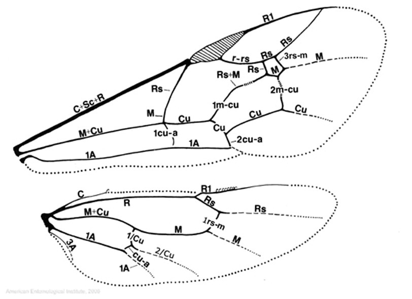
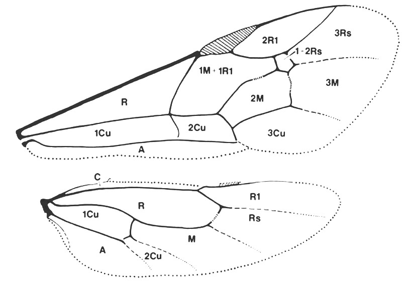
FORE AND HIND WINGS OF AN ICHNEUMONID
(Ross system veins)
FORE AND HIND WINGS OF AN ICHNEUMONID
(Ross system wing cells)
FIRST AND SECOND METASOMAL SEGMENTS, LATERAL VIEW
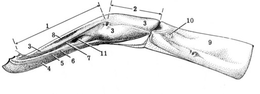
|
1. Petiole 2. Postpetiole 3. Tergite 1 4. Sternite 1 5. Tergosternal suture 6. Ventrolateral carina |
7. Dorsolateral carina 8. Median dorsal carina 9. Tergite 2 10. Thyridium 11. Glymma |
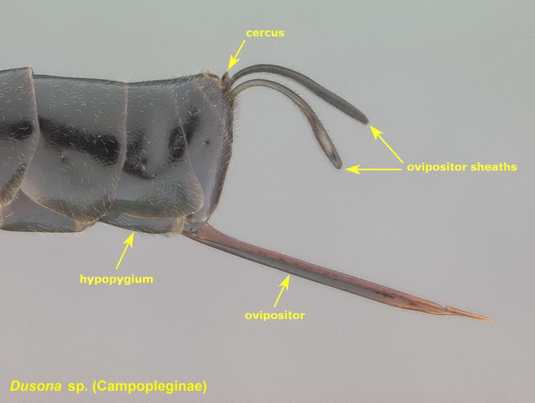
POSTERIOR REGION OF FEMALE METASOMA, LATERAL VIEW
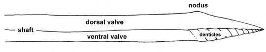
ICHNEUMONID OVIPOSITOR, LATERAL VIEW OF APICAL REGION
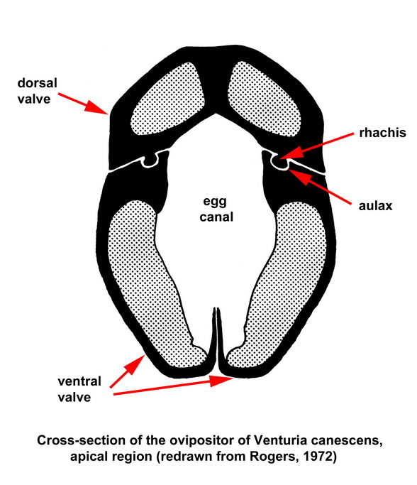
ICHNEUMONID OVIPOSITOR, CROSS-SECTION
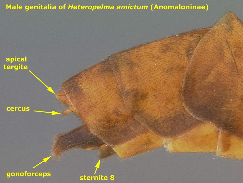
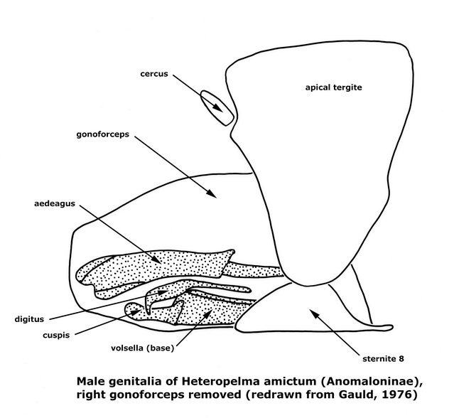
ICHNEUMONID MALE GENITALIA
HEAD
Vertex. The top part of the head, between the inner margins of the compound eyes. It is delimited anteriorly by the anterior ocellus and posteriorly by the occipital carina.
Ocellar triangle. The elevated triangular area containing the three ocelli.
Ocellocular distance (abbreviation: OOD). The shortest distance between the lateral ocellus and the margin of the compound eye, divided by the greatest transverse width of the lateral ocellus.
Supra-antennal area. Part of the anterior aspect of the head, between the eyes, extending from the ventral margin of the median ocellus to the ventral margins of the antennal sockets. Townes and other authors referred to this as the frons.
Supraclypeal area. Part of the anterior aspect of the head, between the eyes, extending from the lower margin of the antennal sockets to the epistomal suture. When the width and height of the supraclypeal area are compared, the width is measured at its narrowest point and height from the anterior tentorial pits to the ventral margins of the antennal sockets. Townes and other authors referred to this as the face.
Malar space. The space between the mandibular socket and the lower edge of the compound eye, sometimes called the cheek in older literature. The length of the malar space is measured from the ventral margin of the compound eye to the mandibular socket, at its narrowest point.
Subocular groove. A groove (occasionally weak and indicated only by granulate sculpture) located in the center of the malar space, extending from the ventral margin of the compound eye to the anterior articulation of the mandible.
Epistomal suture. A shallow groove separating the clypeus from the supraclypeal area; it is sometimes called the clypeal suture in the older literature.
Anterior tentorial pit. One of two shallow impressions in the epistomal suture between the clypeus and the supraclypeal area, situated near the side a little mesad of the lower corner of the compound eye. It is sometimes called the clypeal fovea in older literature.
Clypeus. The area on the front aspect of the head, above the mouth opening and below the face, usually separated from the supraclypeal area by the epistomal suture. The length of the clypeus is measured on the midline, from the epistomal suture to the ventral margin of the clypeus. The width of the clypeus is measured between its extreme corners at the dorsal sockets of the mandibles.
Labrum . The anterior appendage of the mouthparts, suspended from the clypeus. It is usually concealed by the clypeus; its visibility and shape of the apical margin can be taxonomically useful.
Paraocular area. The area of the head adjacent to the inner margin of the compound eye. Townes and other authors referred to this region as the facial orbit and frontal orbit.
Genal orbit. The region of the gena immediately adjacent to the compound eye.
Gena. The area between the compound eye and the occipital carina. It is delimited dorsally by the vertex and ventrally by the mandibular socket. Townes (1969) referred to this area as the occiput and temple.
Occiput. Part of the posterior aspect of the head, within the occipital carina. Townes (1969) referred to this as the postocciput.
Occipital carina. A subcircular carina on the posterior aspect of the head, between the vertex and posterior margin of the compound eyes and the foramen magnum. Its ventral termination may be adjacent to the posterior articulations of the mandibles, or it may reach the hypostomal carina above the base of the mandible and this can be an important character in Ichneumonidae. The ventral parts are called the genal carinae in the older literature.
Hypostomal carina. One of two carinae on the ventral part of the posterior aspect of head, running from the posterior mandibular articulation to the foramen magnum.
Foramen magnum. The large and approximately circular central hole on the posterior face of the head.
Tyloid. A sensory structure on the flagellomere of a male ichneumonid, usually in the form of a longitudinally elliptic or linear raised area on the outer side of each of several flagellomeres. Tyloids occur usually in males of Cryptinae, Ichhneumoninae, and Diplazontinae, often in males of Orthocentrinae, and rarely in males of Pimpla (Pimplinae). Their shapes and distribution frequently afford useful taxonomic characters. The term has often been used incorrectly for any unusual antennal feature such as the concavities present in male Cylloceria.
MESOSOMA
Mesosoma. The apparent thorax in apocritan Hymenoptera, consisting of the thorax and the fused morphological first segment of the abdomen (the propodeum).
Pronotum. The dorsal sclerite of the prothorax; it is mediodorsally narrow but laterally expanded.
Collar. The more or less elevated anterior margin of the pronotum, just behind the head. The collar crosses the dorsal aspect of the pronotum (where it usually is highest) and extends ventrally and obliquely posteriorly on each side.
Epomia. A carina on the lateral face of the pronotum. Dorsally, it obliquely crosses the shallow depression on the side of the pronotum; it rarely continues ventrally on to and along the anterior pronotal margin.
Tegula. A convex scale lying over the base of the fore wing, on the anterior side.
Notaulus. One of a pair of grooves on the mesoscutum, each beginnning on the anterior margin to one side of the midline and extending posteriorly. The notauli divide the mesoscutum into 3 parts: a median lobe between the two notauli and a lateral lobe on each side.
Scutellum. A median, subtriangular, raised part of the mesonotum, behind the mesoscutum.
Postscutellum. The small median raised part of the metanotum, lying between the apex of the scutellum and the base of the propodeum.
Mesopleuron. It is composed of the mesepisternum and mesepimeron. The mesepisternum is anterior the mesopleural suture; it comprises most of the mesopleuron in ichneumonids and is the structure termed mesopleuron in descriptive taxonomy. The mesepimeron is posterior of the pleural suture; it is a narrow band that usually is ignored when describing the mesopleuron.
Mesopleural groove. A vertical or somewhat oblique groove near the hind edge of the mesopleuron, reaching from the near the middle coxa to the base of the front wing. It separates the mesepisternum from the mesepimeron.
Epicnemial carina. A carina paralleling the anterior margin of the mesepisternum and delimits the posterior margin of the epicnemium. The epicnemial carina often meet ventrally, thus presenting a single transverse carina on the anterior part of the mesothoracic venter. Townes referred to this as the prepectal carina.
Epicnemium. The area along the anterior margin of the mesepisternum, delimited posteriorly by the epicnemial carina.
Subalar ridge. A transverse ridge near the dorsal edge of the mesopleuron, beneath the tegula and base of the fore wing. It is sometimes called the subtegular ridge in the older literature.
Hypoepimeron. A weakly raised, polished or less strongly sculptured area on the dorsal posterior part of the mesepisternum. It is sometimes called the speculum by some authors.
Scrobe. A pit or a short horizontal groove on the mesopleuron, at its midheight, just in front of the mesopleural groove and below the hypoepimeron. It is sometimes called the mesopleural fovea in older literature.
Sternaulus. A horizontal groove on the ventral region of the mesopleuron, starting at the epicnemial carina at the level of the fore coxa and extending posteriorly, sometimes reaching the base of the middle coxa. The sternaulus is considered the dividing line between the mesopleuron and the mesothoracic venter. It is not to be confused with the long foveate groove found in tersilochines and some other scattered taxa: the foveate groove starts at the epicnemial carina at about the midheight of the mesopleuron, and extends ventrally to near the middle coxa. The sternaulus is more or less level, while the foveate groove is at a 20°-40° angle.
Posterior transverse carina of the mesothoracic venter. A transverse carina on the ventral surface of the mesothorax, just in front of the middle coxae. The carina may be complete, may be interrupted in front of each of the middle coxae, or may be obsolescent with only a short trace on each side near the coxal socket and perhaps a remnant at the midline. This carina was called the postpectal carina by Townes.
Metapleuron. The metapleuron is in two distinct parts. The ventral division is the larger, and ordinarily is the only part referred to when the metapleuron is mentioned. This ventral part is an oval or subtriangular area on the side of the mesosoma, between the middle and hind coxae and extending up to the propodeum. The dorsal division of the metapleuron lies behind the upper half of the mesepimeron and below and a little behind the base of the hind wing. It is separated from the propodeum by a furrow.
Submetapleural carina. A carina on the ventral margin of the ventral division of the metapleuron, between the bases of the middle and hind coxae.
Juxtacoxal carina. This carina is on the ventral division of the metapleuron, originating from the anterior part of the submetapleural carina and extending dorsally.
Propodeal furrow. The deep groove found between the metanotum and propodeum, present in almost all ichneumonids. It incorporates the metapostnotum (Wahl, 1985).
Propodeum. This is the first abdominal segment of apocritans that has become fused to the thorax. It bears a number of carinae that delimit various areas. These are quite important for ichneumonid taxonomy.
Propodeal carinae:
Anterior transverse carina. The anterior one of the two transverse carinae of the propodeum. There is sometimes reference to the costula: this is the sublateral section of the anterior transverse carina, separating the first from the second lateral area.
Posterior transverse carina. The posterior one of the two transverse carinae of the propodeum.
Lateral longitudinal carinae. The longitudinal carinae of the propodeum are between the median longitudinal and pleural carinae. There is one lateral longitudinal carina on each side.
Median longitudinal carinae. The median pair of longitudinal carinae of the propodeum.
Pleural carina. The carina between the propodeum and metapleuron (ventral division of the metapleuron).
Propodeal areas:
Basal area, or median basal area. The median area at the base of the propodeum, bounded on each side by the median longitudinal carinae and posteriorly by the anterior transverse carina. This is the area basalis of European authors.
Areola. The median area on the propodeum, usually pentagonal or hexagonal in shape. The areola is bounded laterally by the median longitudinal carina, anteriorly by the anterior transverse carina, and posteriorly by the posterior transverse carina. This is the area superomedia of European authors.
Petiolar area. The median region of the propodeum just above the metasomal attachment, bounded dorsally by the posterior transverse carina. When the posterior transverse carina is absent (as is often the case), the areola and petiolar area are said to be confluent. This is the area petiolaris of European authors.
Lateral areas. The propodeal region between the median longitudinal and lateral longitudinal carinae is divided into three parts by the transverse carinae (when present): the first, second, and third lateral areas. They are referred to by European authors as (respectively) area externa, area dentipara, and area postero-externa.
Pleural areas. The propodeal region between the lateral longitudinal carina and the pleural carina is divided into three parts by the transverse carinae (when present): the first, second, and third pleural areas. They are referred to by European authors as (respectively) area spiracularis, area lateralis, and area juxta-coxal. The propodeal spiracle is located in the first pleural area. The first and second pleural areas are often confluent.
Propodeal apophyses (singular: apophysis). Two teeth, horns, crests, or tubercles located on each side of the propodeum near its midlength, situated at the hind end of the second lateral area or sublaterally on the posterior transverse carina.
Propodeal spiracle. Measurements of the length and width are made to the edge of the surrounding rim. The opening itself is not the thing measured.
Trochanter and trochantellus. In ichneumonids there are usually small segments between the coxa and the femur. The anterior one is called the trochanter. Posterior to this is the trochanatellus, which is derived from the basal end of the femur. In a few ichneumonids, the trochantellus is not separated from the femur.
Pectinate This term is applied to tarsal claws that have a series of small teeth on the ventral margin. The teeth may be triangular, or so elongate and close together that the claw appears like a comb. All, or nearly all, tarsal claws of ichneumonids are in fact pectinate, but often the teeth are few, crowded to the base of the claw, and so inconspicuous that a microoscope slide preparation is required to show them clearly. The tarsal claws are said to be not pectinate or "simple" when no pecten teeth can clearly be seen without a microscope slide mount.
WINGS
Wing veins. The Comstock and Needham system of vein terminology, as modified by Ross (1936) and Mason (1990), is used here. Veins are divided into abcissae, or sections, by their intersection with other veins. For example, the second abcissa of fore wing vein 1A (between 1cu-a and 2cu-a) is written as "2/1A." Measurements of the length of wing veins or sections of wing veins is made from the center of one juncture to the center of the next. Mason (1986) recognizes four types of wing veins:
tubular: a rigid tubular structure with sharply defined edges; the normal wing vein of Hymenoptera.
nebulous: a more or less pigmented vein without a tubular structure, thus having ill-defined edges; appear darkest centrally and fade gradually toward the edges.
spectral: an unpigmented vein that is normally invisible by transmitted light but can be seen by light reflected off the wing membrane because its course is marked by a furrow or ridge on the wing surface.
adventitious: markings appearing like nebulous or even tubular veins but occur in places where they cannot be homologized with the normal venation.
Wing cells. Wing cell terminology follows a modified Comstock and Needham system (Goulet & Huber, 1993).
Bulla. An unpigmented section of a wing vein, marking the area where a fold or flexion line of the wing crosses the vein.
Inclivous/vertical/reclivous. Terms applied to the orientation of a transverse wing vein. Inclivous means that its anterior end is nearer the wing base than its posterior end. If both ends are equally distant from the wing base, the vein is vertical; if the anterior end is farther from the wing base than the posterior end, the vein is reclivous.
Areolet. A small cell in the center of the front wing, between veins 2/Rs and 3rs-m (the intercubital veins of some authors).
Stigma. A strongly sclerotized thickened area at the apex of the costal vein on the anterior margin of the fore wing. The shape and color (usually black to dark brown) are often taxonomically valuable.
Hamulus. A small, bristle-like hook on the anterior margin of the hind wing that hooks into a channel on the posterior margin edge of the fore wing to join the two wings together. There are two clusters of hamuli: The distal hamuli number about 10, more or less, and are situated just distad of the base of vein R1. The basal hamuli are on the apex of vein C, number one to about six (usually one), and are often weak or absent. In describing the hamuli, the basal cluster is often ignored and the remarks refer only to the distal hamuli.
METASOMA
Metasoma. The apparent abdomen in apocritan Hymenoptera, not including the morphological first segment which has transferred to the thorax as the propodeum. Metasomal segments in ichneumonids are numbered from anterior to posterior.
First metasomal tergite (also referred to as tergite 1 or T1). When the length and width of this sclerite is compared, the length is measured from the center of the insertion area of the extensor tendon to the extreme apex on the midline. (The extensor tendon inserts on the midline on the dorsal side, near the base of the tergite.) The width of tergite 1 is measured at its widest point, which is nearly always close to the apex. Tergite 1 typically has three pairs of longitudinal carinae: The median dorsal carinae are a pair on the dorsal surface, between the spiracles. The dorsolateral carinae (one on each side) are in the position that the name implies, at the level of the spiracle. The ventrolateral carinae follow the ventral edges of the tergite.
Glymma. A groove or pit on the side of tergite 1, between its spiracle and base. For some unknown reason, the glymma is nearly always present when the sternite is free from its tergite, and absent when the sternite is fused with its tergite. This is therefore a convenient way of determining whether the sternite is free (glymma present) or fused (glymma absent).
Petiolate first metasomal segment. The definition of petiolate is somewhat subjective, but one that was coined at an April 2006 meeting of Andrew Bennett, Jacques Dubois, Ian Gauld, Michael Sharkey, and David Wahl is used here: anteriorly parallel-sided and usually abruptly broadened beyond the spiracles; if the first metasomal segment is parallel-sided throughout its length, then more or less cylindrical and > 2x as long as apical width. The corresponding definition of non-petiolate is: more or less evenly broadened from near base to apical margin; if the first metasomal segment is parallel-sided throughout its length, then clearly depressed and < 2x as long as apical width. If the first metasomal segment is petiolate as defined above, then the petiole is the part of the first metasomal segment in front of the spiracle, and the postpetiole is the widened hind part posterior to the spiracle.
Thyridium [plural: thyridia]. A scar-like area on each side of tergite II, between the anterior margin and the spiracle. It has a different surface sculpture than the rest of the tergite, lacks setiferous punctures, usually has a matte surface, and is often slightly depressed. Its shape varies from roughly circular to transversely ovoid/linear to longitudinally linear. It is found only in the Ichneumonidae (lacking in the Agriotypinae, Anomaloninae, Metopiinae, Orthopelmatinae, and Xoridinae) and certain polistine Vespidae. This is not be confused with the pseudothyridium.
Gastrocoelus. An impunctate impression on each side of the second tergite, near the base. It includes the thyridium, which forms the posterior margin. Gastrocoeli are found only in the Ichneumoninae. The plesiomorphic condition is a relatively deep impression but it has been modified in many ichneumonine lineages, ranging from a shallow depression to a flat and elongate region.
Pseudothyridium. A scar-like ovoid area on each side of tergite II and following tergites, located between the spiracle and the tergal posterior margin. It is present in addition to the thyridium. Whilst thyridia are restricted to Ichneumonidae, pseudothyridia are widely distributed throughout the Hymenoptera.
Hypopygium. The last visible sternite, just in front of the genitalia of the male or the ovipositor of the female. This is metasomal sternite 8 in males and metasomal sternite 6 in females.
Cerci (singular: cercus). A pair of small finger-like sensory organs at the apex of the last metasomal tergite.
Laterotergite. The thin lateral margin of the second and following metasomal tergites. Laterotergites may be completely continuous with the tergite or there may be a horizontal crease or groove that marks the division between the tergite and laterotergite. They may hang down or may be turned mesad to cover the lateral part of the underside of the metasoma. They may also be sharply inflexed against the overhanging edge of the tergite. In such cases they are usually very narrow or obsolescent.
Ovipositor. The ovipositor shaft consists of three sections. The dorsal valve is composed of the two fused second gonapophyses, whilst the ventral valve consists of the two separate first gonapophyses. The ventral margins of the dorsal valve have ridges (the rhachis) which fit into grooves (the aulax) on the dorsal margins of the ventral valve, thus locking the three elements together into a hollow tube through which the egg passes. The apex of the dorsal valve may have denticles/ridges, or may simply be smooth; the apex of the ventral valve usually has a series of oblique denticles/ridges. The ovipositor shaft has two paired covering parts, the ovipositor sheaths (= gonoplacs). The length of the ovipositor sheaths, in relation to the length of the hind tibia or of the fore wing, is often used to indicate the length of the exserted part of the ovipositor, as the lengths of the sheaths and exserted part of the ovipositor are the same.
Nodus. A dorsal prominence on the tip of the ovipositor, a short distance before the apex.
Gonoforceps (singular and plural). The paired lateral outer parts of the male genitalia. The gonoforceps is probably a fusion of the gonocoxite and gonostylus. Other names for the structures are gonosquamae or parameres or "claspers".
Aedeagus. The sclerotized apical section of the intromittent organ, resulting from the fusion of the gonapophyses of abdominal segment 9. Other names are penis, penis and penis valves, and phallus.
Volsellae. [singular: volsella]. A pair of lateral processes that flank the aedeagus. Each volsella projects internally from the gonoforceps; its lateral edge is attached to the gonoforceps. At the apex of the volsella is an interior moveable digitus (= gonolacinia) and an exterior immovable cuspis (= distivolsella); these form a pincer and are used to grasp the female during coition.
References
Bohart, R.M. & A.S. Menke. 1976. Sphecid Wasps of the World. University of California Press: Berkeley. 695 pp.
Gauld, I.D. 1976. The classification of the Anomaloninae (Hymenoptera: Ichneumonidae). Bulletin of the British Museum (Natural History), Entomology 33: 1-135.
Huber, J.T. & M.J. Sharkey. 1993. Chapter 3 Structure. In: Goulet, H. & J.T. Huber, J.T. (eds.). Hymenoptera of the World: An Identification Guide to Families. Ottawa: Agriculture Canada Publications, Ottawa. 668 pp.
Mason, W.R.M. 1986. Standard drawing conventions and definitions for venational and other features of wings of Hymenoptera. Proceedings of the
Entomological Society of Washington, 88: 1-7.
Mason, W.R.M. 1990. Cubitus posterior in Hymenoptera. Proceedings of the Entomological Society of Washington, 92: 93-97.
Michener, C.D. 1944. Comparative external morphology, phylogeny, and a classification of the bees (Hymenoptera). Bulletin of the American Museum
of Natural History 82: 151-326.
Rogers, D. 1972. The Ichneumon wasp Venturia canescens: oviposition and avoidance of superparasitism. Entomologia Experimentalis et Applicata 15: 190-194.
Ross, H.H. 1936. The ancestry and wing venation of the Hymenoptera. Annals of the Entomological Society of America, 29: 99-111.
Townes, H. 1969. The genera of Ichneumonidae, part 1. Memoirs of the American Entomological Institute, 11: 1-300.
Wahl, D.B. 1985. A revision of the genus Agathilla (Hymenoptera: Ichneumonidae). Transactions of the American Entomological Society, 111: 267-277.
Site credits and citation. Questions? E-mail: aei[at]aei.cfcoxmail.com